|
Today, the day after the surgery, we still aren't sure what caused all this. The problem he had was a detached retina of the left eye, which caused him to lose sight in that eye. An infection started as well. It isn't clear right now if the detached retina caused the infection, or the infection caused the detachment. The vet sent the removed eye to a pathology lab where they hope to be able to figure out the sequence of events. Two Wednesdays ago we noticed the area around his eye was a little bit red. Nobody thought much about it then. The next morning, it was still red and started to swell up a little bit; by the afternoon, it was very swollen. We took him to the vet. The vet was stumped. Searched all around the eye for a foreign body. Didn't find anything. He diagnosed Charlie with conjunctivitis (pink eye). We got some steroid eye drops and were told to come back Monday if things weren't better. This all seemed very reasonable. Nobody figured anything major was wrong. Sunday, the eye was even more cloudy. You
couldn't really see any of the brown anymore. The swelling was down a bit
because of the steroids,
but it obviously was still bothering him and looked different, but worse.
Monday morning, the eye was blue and all the way clouded over. He was clearly photo-sensitive in that eye and kept it close most of the time. We took him to a doggie ophthalmologist. We were getting very worried and wanted a specialist. He also was stumped. No obvious explanation. Said bring him back tomorrow morning (Tuesday) at 7:00am and they will do some tests; but said the prognosis probably wasn't good. They took blood, did an aspirate (withdrew liquid from the eye), and an ultrasound of the eye. The ultrasound revealed the detached retina causing Charlie to loose his site in that eye. There is no recovery from that - detached = no vision. On Wednesday some of the lab results started returning, and confirmed an infection. We still hadn't identified what caused all this,
and were all (vets included) very concerned. We all feared some big
problem. As more lab results came in, we were asked to take him back to
the normal vet for a chest x-ray. Apparently one of the lab tests found
some fungus in the eye along with the infection. The ophthalmologist was
worried the
Only solution to a problem like this is to have the eye removed. Once vision is gone, there isn't any real reason to leave the eye in, and the infection was making it important to get the problem out of there. This visit and realization came on Thursday - 8 days after the original redness. He was figured to have been fully blind in that eye only 5 days after we first noticed the redness. We scheduled the surgery to remove the eye the first available time the ophthalmologist had - which was the following Wednesday. Leading up to the surgery day, we had to decide
how to handle it - just sew the eye shut, sew it shut with a ball in the socket
(to make it look more natural), or to get a fake eye. The fake eye option
was the source of much discussion. Apparently they leave the very front
layer of the normal eye in place, and put the fake eye in behind it. So
the thin eye layer is holding the fake in place. Advantages of this are he
looks more natural - but there is some small chance of complications in the
future. I was really worried about the fake eye looking creepy, but Renee
was leaning towards getting the fake eye as a better option to keep his beautiful
face unchanged.
Wednesday the 28th rolls around - finally. Surgery day. 7:00am we drop him off. Renee picked him up at about 10. He did excellent. No complications, nothing unusual. He was pretty groggy all day, whimpering quite a bit around the house that first day. The vet said his whimpering was probably just being confused from the surgery and drugs rather than pain. They pumped so much Novocain into the eye socket, he said Charlie wouldn't feel anything in there for 48 hours. He slept the entire night through, which is a good sign. The vet called us to check on him Wednesday afternoon, and again Thursday. They seem to really care about him. They sewed the eye closed, and then sewed a big piece of gauze over the eye, so we can't really see what he will look like yet. The gauze comes off next Wednesday, and the underlying stitches the following week. After two weeks, he should be fully recovered and good-to-go back to normal activities. In the meantime he is wearing an Elizabethian collar (a satellite dish) around his neck to keep him from scratching at the eye. He doesn't like the dish at all.
We are just so happy the problem was only his eye and nothing more major. He is a great dog, and we love him so much. While it's agonizing to have something like this happen, we are glad we are in a position to get him the good care and make him better. Below is some information about retinal detachment I found on the web. It does a pretty good job describing the situation. "Retinal detachment means that the sensory retina has separated from the back of the eye. The retina lines the back of the inner eve and contains the light-sensitive rods and cones that change light into energy for transmitting messages to the brain. The retina is similar to the film in a camera--the image or picture is received on it.
For some types of retinal detachments, a combination of surgeries is performed. Unfortunately, retinal detachment surgery is not always successful. Chronic retinal detachments can lead to intraocular bleeding and may increase the risk for developing glaucoma. Otherwise, a retinal detachment is not painful."
|
 Wednesday April 27, 2005 Charlie had
his left eye removed. Charlie is our black lab, about 18 months
old when he had to have the eye taken out. Very sad, but the
vets all say he will be fine, adjust to it, and live a full happy
life without it.
Wednesday April 27, 2005 Charlie had
his left eye removed. Charlie is our black lab, about 18 months
old when he had to have the eye taken out. Very sad, but the
vets all say he will be fine, adjust to it, and live a full happy
life without it. We
decided not to wait until Monday, and took him back to the vet. A
different vet (at the same office) saw him. She was stumped as well.
Gave us some different antibiotic eye drops and said bring him back on Tuesday if things
aren't getting better.
We
decided not to wait until Monday, and took him back to the vet. A
different vet (at the same office) saw him. She was stumped as well.
Gave us some different antibiotic eye drops and said bring him back on Tuesday if things
aren't getting better.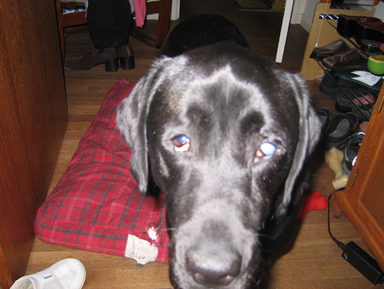 fungus
may have been inhaled and may be in his lungs, which would be very bad. X-rays were fine.
No lung problems. Blood tests were fine - overall good health.
Everything was localized to the eye problem - the detached retina and the
infection there.
fungus
may have been inhaled and may be in his lungs, which would be very bad. X-rays were fine.
No lung problems. Blood tests were fine - overall good health.
Everything was localized to the eye problem - the detached retina and the
infection there. We
both flip-flopped our position over the next few days. In the end, we
didn't have a real decision - the vet said with the infection, it wouldn't be
wise to do the fake eye. So we easily agreed to sewing it closed with the
silicon ball in the socket to make it look more natural (otherwise it would be
sunken in a little bit without anything behind the skin).
We
both flip-flopped our position over the next few days. In the end, we
didn't have a real decision - the vet said with the infection, it wouldn't be
wise to do the fake eye. So we easily agreed to sewing it closed with the
silicon ball in the socket to make it look more natural (otherwise it would be
sunken in a little bit without anything behind the skin). The vet put a big sticker on the collar that says
"I was very brave". He certainly has been a trooper. They say he
will fully be fine without the eye, not even missing it. He will still be
able to catch balls, run around crazy, and do all the normal dog things.
Dogs are very adaptive. Their eye sight isn't like humans
where the eyes work together to give depth perception. Dogs eyes
work more individually, so missing one just cuts down on the peripheral vision a
bit, not really impacting normal sight.
The vet put a big sticker on the collar that says
"I was very brave". He certainly has been a trooper. They say he
will fully be fine without the eye, not even missing it. He will still be
able to catch balls, run around crazy, and do all the normal dog things.
Dogs are very adaptive. Their eye sight isn't like humans
where the eyes work together to give depth perception. Dogs eyes
work more individually, so missing one just cuts down on the peripheral vision a
bit, not really impacting normal sight.